Monday, January 11, 2016

Key Givens


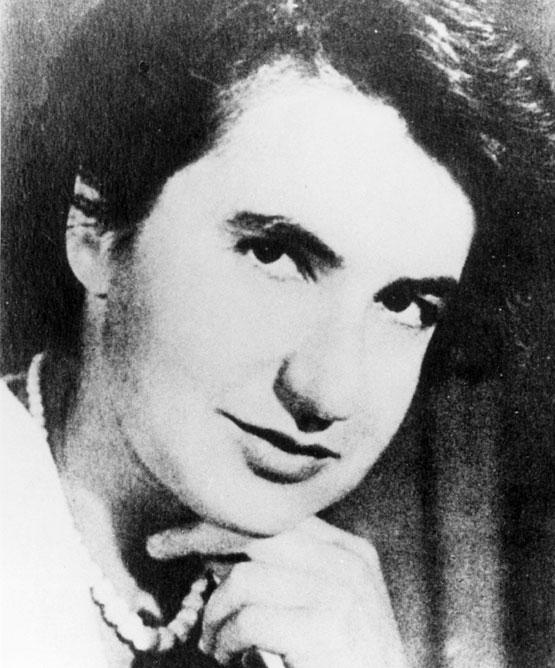
  • Rosalind Frankin - 30's. Jewish.
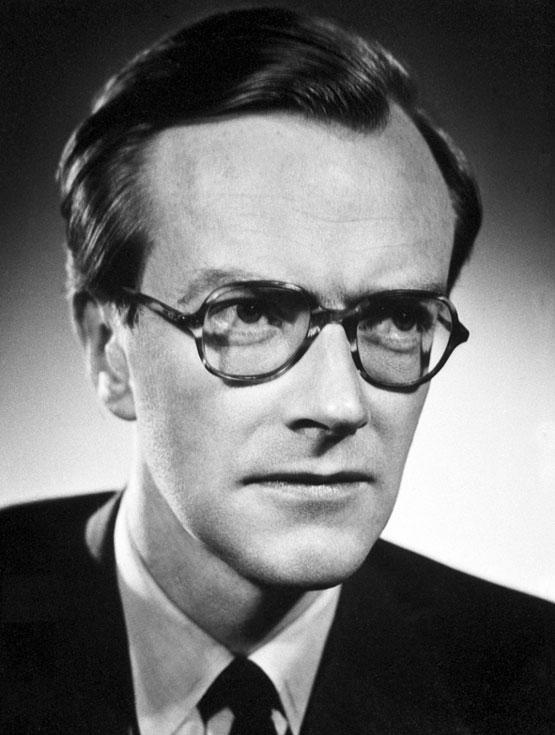
  • Maurice Wilkins - 30's/40's.
  • Ray Gosling - 20'.
  • James Watson - Early 20's.
  • Francis Crick - 30's/40's. His hair is amazing/crazy.
  • Randall, aka Sir John Turton Randall
         b. 1905 (46 - 48 years of age). Led the King's College London team. Wilkins is his deputy.
  • Linus Pauling
         
    (February 28, 1901 – August 19, 1994)[4] was an American chemist, biochemist, peace activist, author, and educator. He published more than 1200 papers and books, of which about 850 dealt with scientific topics.[5] New Scientist called him one of the 20 greatest scientists of all time,[6] and as of 2000, he was rated the 16th most important scientist in history.[7] 
    For his scientific work, Pauling was awarded the Nobel Prize in Chemistry in 1954. Pauling also worked on DNA's structure, a problem which was solved by James Watson, Francis Crick, Rosalind Franklin and Maurice Wilkins.[10]
  • William Bragg 
         (31 March 1890 – 1 July 1971) was an Australian-born British physicist and X-ray crystallographer, discoverer (1912) of Bragg's law of X-ray diffraction, which is basic for the determination of crystal structure. He was joint winner (with his father, William Henry Bragg) of the Nobel Prize in Physics in 1915: "For their services in the analysis of crystal structure by means of X-ray",[3] an important step in the development of X-ray crystallography.
    Bragg was knighted in 1941.[4] As of 2016, Lawrence Bragg is the youngest ever Nobel Laureate in physics, having received the award at the age of 25 years.[5]  Bragg was the director of the Cavendish Laboratory, Cambridge, when the discovery of the structure of DNA was reported by James D. Watson and Francis Crick in February 1953.
  • Odile Crick.  Wife of Francis Crick.

Takes place between 1951 & 1953.

It is "particularly cold in London, January 1951" - Wilkins

Wilkins & Crick = BFFs.
Crick's new co-worker is Watson, who Wilkins also has a work crush on.

"The Jews can be very ornery." - Watson

Behind her back, the doctors call her Rosy.

Watson says Rosalind's handshake is too firm, that there is nothing gentle, nothing remotely tender about her.

Wilkins, of the U.S., "theft and burglary are upheld as virtues", it's "turning its sinners into saints".

Wilkins + wife + son = :(

Wilkins is "unhappy" about Don's arrival.

Wilkins says Rosalind has been distracted since Don's arrived. Don says she was not.

Watson & Crick's new conclusion is that DNA consisted of TWO chains running in opposite directions, a pair of endless spirals that will work together but will never meet. This is how it replicates. How it works. Each strand is a template and in each template is another helix and on and on forever.

Mid-February, when they are close to cracking the secret of life, Watson and Crick invite everyone but Gosling to Cambridge. They are particularly cheerful. Everyone can see that Don is in love with Rosalind at this event.

Watson and Crick struggled with how the 4 bases fit into the picture. Did they pair? Work together? Or were they distinct from each other?

February 28, 1953 is the evening Watson and Crick crack the secret and the same evening Don and Rosalind go on their date to an Italian restaurant. Rosalind is taken to the hospital and diagnosed with ovarian tumors later that evening.

From 1956 onwards, Franklin battled with numerous tumors - possibly due to her work with radiation sources in an era before they were at their safest. She took ill and died April 16, 1958 from secondary illness related to her ovarian cancer. It was only after her death that allegations of sexism began to arise. As Crick and Watson's fame grew for solving the great "mystery of life," Franklin's contribution became difficult to ignore - although it would have been difficult to properly cite it in the literature anyway, as Franklin had mostly kept Photo 51 hidden from Wilkins and never published it.

Don Caspar Thoughts

Don Caspar Notes

Don Caspar - American, Jewish. 20's/30's. "A doctoral student in biophysics at Yale, close to getting PHD" (Gosling). His advisor is Simon Dewhurst.
He is considering the chemical makeup of coal as a suitable topic for the final stage of his doctoral research.** He is looking to combine an applied and theoretical approach. 
**Coal is defined as a readily combustible rock containing more than 50% by weight of carbon. Coals other constituents include hydrogen, oxygen, nitrogen, ash, and sulfur. Some of the undesirable chemical constituents include chorine and sodium.


A) Coal originates from vegetal matter and forms from the compression of peat over millions of years (Figure 1).


Figure 1: Formation of Coal
  Source: Coal Exploration, Mine Planning, and Development

B) It is a stratified, organic, biochemical sedimentary rock.
C) Coal is a heterogeneous mixture of plant materials and minerals.
        -Ex. wood, bark, leaves and sap.
D) The calorific value (which is the calories or thermal units contained in one unit of a substance and released when the substance is burned) of coal and lignite varies tremendously; ranging from 15,000 BTUs/lbs to under 6,300 BTUs/lbs.  These values, however, are the extremes.  Most ranks of coal are between these two levels.
E) The organic component of coal consists of chemical compounds from carbon, hydrogen, oxygen, sulfur, and nitrogen. (Figure 2)


Figure 2: Composition of Coal
             Source: www.btinternet.com/~ian.rivett/ imic/combust.htm            

F) With the exception of nitrogen, these elements are also found in the mineral species that occur in coal.
         - Ex. carbonates, sulfides, sulfates and hydrous clay minerals.
         - The ash in coal is made of decomposed residues of silicate,     
          carbonates and sulfides.
  
G) Coal is ranked in six different categories:
 1. Peat - not really a coal; it is undeveloped, soft and spongy. It is 20-25% carbon, and was deposited during the last 100 years by decomposed plant remains.    
 2. Lignite- the first "real" coal.  It has a low heating value and is often found near the earth's surface. Lignite coal loses its moisture when exposed to air.
 3. Subbituminous - lower in carbon content than bituminous coal.  It also loses its moisture when exposed to air.  When exposed to wetness it
 4. Bituminous - has a high carbon content (65-85%), and a low oxygen content (5-15%).  It usually burns with a yellow flame.
 5. Semianthracite - a hard coal but softer than anthracite.  It has a fixed carbon percentage of 80-85%.  It contains more volatile matter than anthracite and also burns more freely.
 6. Anthracite - hard, durable, high density.  It has low volatile matter and is 85-98% fixed carbon.  It ignites slowly and burns with little flame or smoke.
H) Hydrogen and Carbon Composition
1. Low-rank coals typically have relatively low carbon contents and relatively high oxygen contents (Figure 3).


Figure 3: Percent Hydrogen, Percent Carbon
   Source: Coal Exploration, Mine Planning, and Development

2. Lignite, subbituminous, and low rank bituminous coals have hydrogen    contents of 5-6% (Figure 3).
3. High rank bituminous and anthracite coal are 3-4% hydrogen (Figure 3).

When he graduated, (he says) his parents didn't think he'd ever graduate, neither did he. 

He's had a happy home, parents, & childhood.

Told Dr. Franklin she was "remarkable".

Mid-February, when they are close to cracking the secret of life, Watson and Crick invite everyone but Gosling to Cambridge. They are particularly cheerful. Everyone can see that Don is in love with Rosalind at this event.

Rosalind doesn't mind the way Don calls her "Rosy".

Don's fellowship is only scheduled to run a few months past February (May-Juneish)

He says Rosy was not distracted by his being there.

They go to the garden together.
     Hey Rosy, this sure is a lovely garden. Say, I've been thinking and, well, gosh, why not use ISOMORPHOUS REPLACEMENT with the TOBACCO MOSAIC VIRUS? If you put atoms of lead, or maybe mercury - something heavy - into the virus protein to see what the difference would be between X-RAY patterns. The X-RAY with the atoms would retain the shape of the virus, as opposed to the atoms that would not. That would determine the structure.


Terms Explained

Photograph 51: 
The nickname given to an x-ray diffraction image of DNA taken by Raymond Gosling in May 1952, working as a PHD student under the supervision of Rosalind Franklin at King's College, London in Sir John Randall's group. It was critical evidence in identifying the structure of DNA.

**It confirm the double helical/ anti-parallel structure of DNA**


Cavendish Laboratory:The Cavendish Laboratory is the Department of Physics at the University of Cambridge, and is part of the School of Physical Sciences. The laboratory was opened in 1874 on the New Museums Site as a laboratory for experimental physics.

https://en.wikipedia.org/wiki/Cavendish_Laboratory

DNA: "Nucleic acid is a macromolecule composes of chains of mono-numeric nucleotides. This nucleic acid, or DNA, exists in 2 forms -


an A form and a B form (and now, we know, a Z form)." - Dr. Rosalind Franklin
DNA absorbs lots of water (H2O)

PHOSPHATES:
The phosphates exist on the outside of the molecule.

A phosphate group consists of a central phosphorous surrounded by four oxygens.
The phosphorous is single-bonded to three of the oxygens and double-bonded to the fourth. Due to the nature of the chemical bonds, there is a negative charge on each oxygen that has only one bond coming off of it. This negative charge accounts for the overall negative charge on the phosphate backbone of a DNA molecule.
Phosphates 
can carry energy, shape your cell membranes, and help form the backbone of your DNA


AGCT:
(A) adenine (larger)
(G) guanine (larger)
(C) cytosine (smaller)
(T) thymine (smaller)

(A) pairs with (T). (G) pairs with (C).

CYCLOTRON
cyclotron is a type of particle accelerator invented by Ernest O. Lawrence in 1932 in which charged particles accelerate outwards from the centre along a spiral path. The particles are held to a spiral trajectory by a static magnetic field and accelerated by a rapidly varying (radio frequency) electric field.

Suggested Reading


  • The Double Helix by James D. Watkins
  • Run by Ann Patchett
  • The Eighth Day of Creation by Horace Judson
  • A Winter's Tale by William Shakespeare


King's College

DNA: the King's story

Maurice Wilkins, 1962Maurice Wilkins, 1962This exhibition is a celebration of the work of the King's men and women who helped crack the code of DNA. They include physicists, biologists, chemists and mathematicians.
To quote Francis Crick in 2001: "It is important to remember that all the really relevant experimental work on X-ray diffraction patterns of DNA fibres was done by Rosalind Franklin and Maurice Wilkins and their co-workers."
Rosalind FranklinRosalind FranklinThe exhibition demonstrates both the degree of precision needed and the interconnectivity of the sciences in reaching an understanding.
What follows are the earliest and key images of DNA taken at King’s, the equipment used and pictures of the people most involved.
The exhibition was first launched in 2003 to mark the 50th anniversary of the discovery of the double helix structure of DNA. This updated version includes previously unseen photographs and material catalogued as part of the current Wellcome Trust funded project, 'DNA and Social Responsibility'.  A related photostream of DNA and Social Responsibility images is also online now.
Since the exhibition first apeared, the catalogue to the Maurice Wilkins collection at King's College Archives has been completed and placed online as part of a project supported by the Wellcome Trust.
As well, a significant portion of the collection items are now available online in the Maurice Wilkins and Medical Research Council Biophysics Unit archive.  This is part of Codebreakers: Makers of Modern Genetics hosted by the Wellcome Library.
In preparing this exhibition and obtaining permission to use photographs, the staff of Archives at King’s College London would like to acknowledge the generous support of the family of Maurice Wilkins, Raymond Gosling, Churchill College Cambridge, Jennifer Glynn, Seweryn Chomet, the Nobel Foundation and the Cavendish Laboratory, Cambridge.
Found at:
http://www.kingscollections.org/exhibitions/archives/dna/

Signer's DNA

Signer's DNA

Bottles with Signer DNABottles with Signer DNAIn 1950, Rudolf Signer, a Swiss biochemist based at the University of Berne, who had developed techniques of extracting high-quality DNA from cells, came to London to a Faraday Society meeting and distributed samples to research workers who expressed an interest, including Maurice Wilkins.
The DNA was extracted from a calf's thymus gland. Signer's DNA, and that supplied by Hans Schwander enabled experiments to be designed to explore the structure of this very large molecule.
The same year, Randall recruited Alexander Stokes. He was an experienced crystallographer and an excellent mathematician.
Gosling image of herring spermGosling image of herring spermAlso in 1950, Raymond Gosling, a research student, began to study DNA in sperm heads which Randall was investigating using an electron microscope. Gosling used x-ray diffraction and was advised to pass x-rays through hydrogen to produce a more concentrated image than using air.
Wilkins found Signer DNA unique because it could be pulled or spun into thin filaments of remarkable uniformity that he felt might have a very regular structure and therefore give sharp x-ray diffraction patterns.
Wilkins was noted as being especially adroit at spinning out the fibres, and was described by Gosling as 'being like a wonderful spider'. (When Rosalind Franklin used similar DNA, she employed a highly polished and oiled microscope without the lenses to help pull the samples into the long strands necessary for successful photography.)
Sample holder used to photograph DNA at King'sSample holder used to photograph DNA at King'sThe fibres were so fine (10-30 μm) that they couldn’t be seen unless the light was arranged so that it could be reflected.
This not withstanding, Gosling used the samples to provide the first clearly crystalline x-ray diffraction picture of DNA prior to Franklin's arrival. He and Wilkins realised that precision could be improved by use of a microfocus x-ray tube attached to the camera.
One had just been developed by Werner Ehrenburg and W E Spear at Bernal's laboratory at Birkbeck College. Birkbeck gave the equipment to King's which was modified in King's well-equipped workshops.